
< Previous | Contents | Next >
Factors that Determine How Your Smile Looks
A
great looking smile creates an illusion of energy. While it seems that people with attrac- tive and alluring smiles all have big, white, straight teeth, closer examination reveals that people with great looking smiles do not necessarily have the same perfect teeth.
Beautiful teeth come in a wide variety of shapes, sizes, arrangements and colors that suit each person’s face individually.
Like a fine oil painting, each smile is a canvas containing the application of many elements, and the unique relationship of these elements to each other. Just as an artist combines elements to create a painting that is pleasing to the eye, a cosmetic dentist chooses elements for teeth that will produce the perfect smile. Just as no two paintings are alike, no two smiles are the same.
Many choices and variations must be consid- ered in creating a beautiful smile. The patient’s own perception of beauty must be taken into consideration. By combining the art and science of smile design with the patient’s desires, an aesthetically pleasing smile can be created. Although science dictates the methodology used, the procedure is an artistic creation. The process is a collaboration between doctor and patient. In order for the process of smile creation to be suc-
Factors that Determine How Your Smile Looks

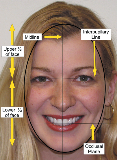
figure 1
cessful, it is absolutely essential that excellent communication exists between the cosmetic den- tist and the patient.
Average patients usually have little or no knowledge about cosmetic dentistry. They may know what they like or don’t like, but they may not know why. Here are the basics of cosmetic dentistry and the principles that guide visual appearance.
Everyone possesses certain basic facial characteristics. There are specific fundamentals that apply to the human head. For example, the human head is oval. The eyes are located on a horizontal plane that bisects the head into two halves; the upper half and the lower half (figure 1). This bisecting line, called the interpupillary line, connects the pupils of the eyes. The interpupillary line connecting the centers of the eyes establish- es a reference point for the horizontal center of the face. Other facial features, including teeth, are balanced with it. When viewed from the front of a face the horizontal plane of the teeth line up parallel to this line. This parallel alignment is known as the frontal view occlusal plane or plane of occlusion of the teeth.
The most important vertical line on the face is the midline, which divides the face vertically into two halves. The center of the two front, upper teeth (the upper incisors) most commonly lines up with the midline of the face.
CHAPTER TWO
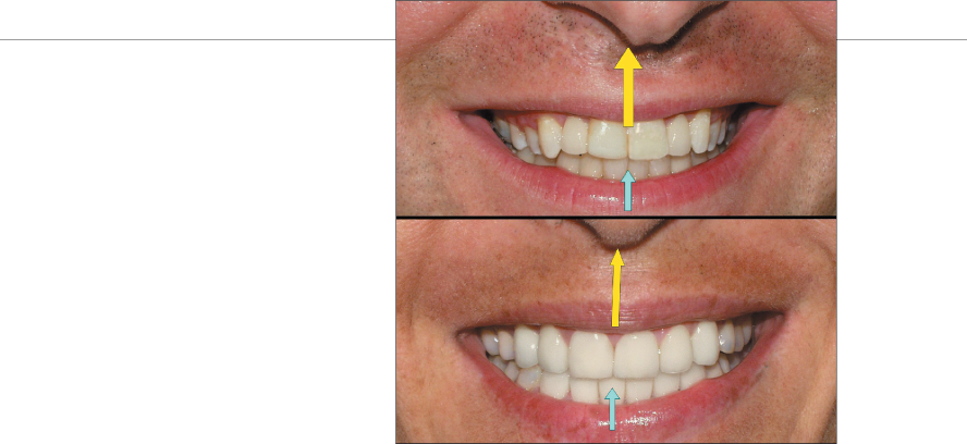
The nose plays an important part in the creative decision making process. When the nose is crooked or off to one side, the decision needs to be made whether the center of the teeth look better centered with the midline of the face or centered with the bottom of the base of the nose (figure 2) Both situations can work on different peo- ple. It’s up to patients to decide what better suits them.
Many times the teeth are the correct shape, size and color, but the plane of occlusion is off on one side. The plane is either too high or too low, making the teeth appear as though they are slant- ing or running up hill or down hill when observed in a mirror.
To achieve a well balanced looking smile, any discrepancy between the occlusal plane and the interpupillary line must be corrected as much as possible. (figure 3)
If one eye is significantly higher than the other, the teeth must be balanced with the other. Here is where the “art” of cosmetic dentistry comes into play.
figure 2
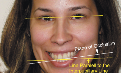
figure 3
Factors that Determine How Your Smile Looks
figure 4
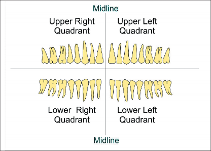
figure 5
If you as a patient want to achieve a partic- ular esthetic look that you believe is correct for your face, you must be able to communicate clearly with your cosmetic dentist about your smile and the teeth within your mouth. In order to do that you must understand how teeth are arranged and what terminology is used to describe them.
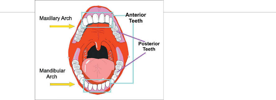
The mouth consists of the upper (maxillary) jaw and the lower (mandibular) jaw. The teeth of each jaw are arranged in an arch and are referred to as the upper (maxillary) arch and the lower (mandibular) arch. The front teeth in both arches from canine to canine (the 3rd tooth from the midline) are called the anterior teeth. All the teeth behind the canines are called the posterior teeth. (figure 4)
When viewing an entire mouth we see that the teeth are also divided into four groups or quadrants. Viewed from the front, they are iden- tified as the upper right quadrant, the upper left quadrant, the lower right quadrant and the lower left quadrant. (figure 5). Using descriptive quadrant terminology helps locate teeth in general areas of the mouth. This quadrant terminology is used when giving feedback to your cosmetic dentist.
CHAPTER TWO

There are 32 individual teeth in the adult dentition. For reference purposes, they are each assigned a number from one to thirty-two (figure 6). The number one tooth starts with the last tooth in the upper right quadrant and continues numeri- cally forward around the arch to the back of the upper left quadrant to the number sixteen. The numbering system continues down to the back of the lower left quadrant to number seventeen and continues forward to the last tooth in the lower right quadrant, number 32. When teeth are entered into a computer or information about them is sent to an insurance company, they are referred to by their numbers. If teeth are missing from the arch, their spaces on the arch are still refered to by their numbers. For example, if tooth number 4 is missing but was going to be replaced by a dental implant, you would say that the implant was being placed in area number 4.
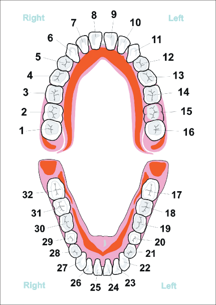
figure 6
Factors that Determine How Your Smile Looks

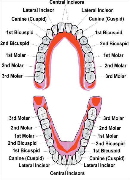
figure 7
Although all teeth are assigned numbers, they are more commonly referred to by their names (figure 7). These names are used by patients and dentists to discuss how they look in the mouth. For more accurate descriptions of a tooth, a location name is used before the name of the tooth is given. For example, when referring to the arch in which teeth are found, the terms max- illa and mandible are used to describe the upper and lower jaws respectively. The first location name is followed by a descriptive second name. The descriptive second name relates to its func- tion in the mouth. The names of the front four teeth are called incisors, so these incisors are referred to as the maxillary and mandibular incisors. The function of the incisors is to tear, penetrate and incise food in the mouth. Incisors are arranged in groups of four. They are the central incisors and the lateral incisors. On each side of the incisors are the canines, (also called cuspids).
After the anterior teeth, (consisting of incisors and canines) are the posterior teeth known as the first bicuspids, second bicuspids, first molars, second molars and third molars (wisdom teeth). As teeth are discussed and described you can see how complex yet accurate this teminology may appear. For example, tooth number 14 is described as the left maxillary first molar. Tooth number twenty-five is the right mandibular central incisor.
This may sound confusing in the beginning, but once the numbers and names become familiar the whole oral naming system will make perfect sense.
CHAPTER TWO

A tooth is much more than a number, location or a name; it is a living entity within the jawbone. It has very specific macro and micro anatomy. The anatomy of a tooth is very com- plex, which is the reason why dentists spend a minimum of four to five years studying dentistry before they have the opportunity to create smiles for patients. For the sake of simplicity, here is a very brief explanation of tooth anatomy. (figure 8)
The part of the tooth that can be seen in the mouth is called the crown. The part that cannot be seen, under the crown and in the jaw bone, is the root. The crown of a tooth consists of a hard outer shell, called the enamel. This is a crystallized structure giving a tooth its hardness, characteristic look and color. Under the enamel is a softer hard layer called the dentin. The color of the dentin may also influence the color of the overlying enamel covering. Within the dentin lies a space occupied by arteries, veins and nerve tissue called the dental pulp, commonly referred to as the nerve. This nerve extends all the way to the bot- tom of the root of the tooth. Each tooth is attached to the surrounding bone by a soft tissue called the periodontal ligament. Covering the bone and surrounding the base of each tooth is a soft pink tissue called the gingiva (commonly known as the gums).
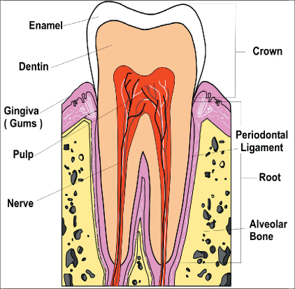
figure 8
Factors that Determine How Your Smile Looks

figure 9
Now that we have a basic understanding of what a tooth is, where it is and what its parts are called, let’s look at the overall smile. (figure 9)
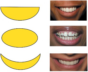
Boat Shape
Oval Shape
Banana Shape
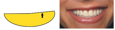
Lip High On One Side
The lips determine the general shape of the mouth when a smile is formed. Some smiles have the shape of a boat, and others look like an oval or a banana. No matter how the teeth are arranged or changed, the lips are the overall cur- tain that frames the teeth and influences the look of a smile. Many times, patients produce pictures, saying, “I want to look like him/her.” The reality is that they can’t. The problem is that these patients do not understand that they have completely dif- ferent shaped lips and mouths. Unless lips, shapes of mouths or arch forms are noted and discussed at the beginning of the consultation with the cosmetic dentist, the patient may never be satisfied. The end result will not meet his/her expectations. And they will never know why.
Lips can also be crooked on one side of the midline of the face and normal on the other. When lips are higher on one side of the midline than on the other, the teeth on the side with the higher lipline will appear larger than the teeth on the other side because more tooth structure shows when the lip is higher. (figure 10)
figure 10
CHAPTER TWO

A very common lip variation is called a “high lip line,” giving a patient a “gummy” smile look (figure11). This occurs when the muscles of the mouth pull the upper lip up too high when smil- ing or speaking or when the bone supporting the upper teeth (maxillary bone) is too prominent. On most people’s smiles, the lips cover the part of the teeth that enters the gum line (called the gingival margins). The actual gum line appear- ance in these patients is not cosmetically impor- tant because it can’t be seen during a smile. When the lip line is high, the gums and shape of the gin- gival contours play a more critical role in how the teeth and smile looks.
Ideally, all the gingival margins should be even, as if they were lined up against an imaginary line at the top of all the teeth. Unfortunately, gingival margins are often at different level giving teeth an unbalanced look or a sense of uneven- ness. Whether it is a single tooth or a group of teeth that have uneven gingival margins, it should be corrected if the gums show when the person smiles. Otherwise, eyes will always focus on the uneven gum line when the person smiles (figure 12). If the gums are covering too much tooth structure, correction can be accomplished by sim- ple surgical procedure such as gingivectomies, crown lengthenings, and gum lifts.
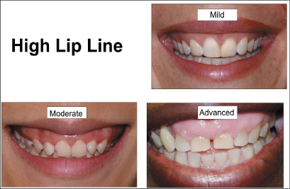
figure 11
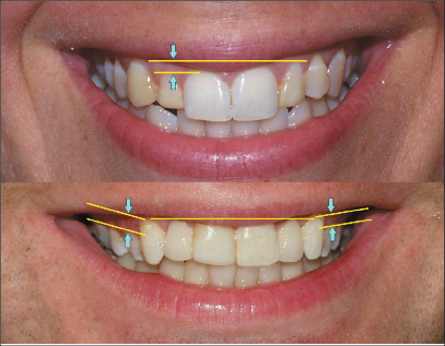
figure 12
Factors that Determine How Your Smile Looks

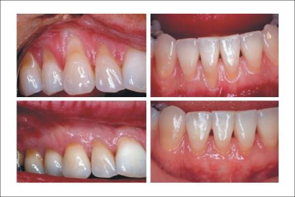
figure 12b
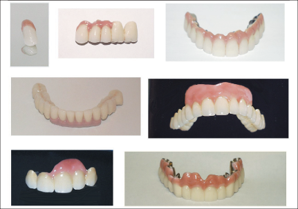
figure 13
Other times the opposite problem exists and there is not enough gum tissue covering the teeth. The gum line appears much too high in the max- illa or too low in the mandible when the patient smiles. This gives the teeth a very long and unnatural look, and they seem out of balance with the oth- ers. If the problem is minimal, a piece of gum tissue can be grafted onto the elongated tooth root recreating a normal appearing tooth. (figure 12b). If the gum line is extremely receded, or the patient is not a candidate for gum grafting (gingi- val grafts), more extensive restorative procedures are performed.For example, teeth can be restored using either porcelain veneers, crowns or fixed bridges, and pink porcelain can be added to these restorations to create the illusion of a natu- ral gum tissue. Thesew types of restorations are particularly useful where teeth have been lost or gums have receded. (figure 13)
CHAPTER TWO
Rounded
Square
Tapered

As mentioned earlier, teeth are arranged on either the maxillary arch or the mandibular arch. The shapes of these arches can vary as well. The arch can be oval or rounded, square or tapered. (figure 14) These individually shaped arches can also be wide, average or narrow.
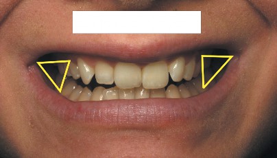
The shape and width of the arch will affect how the teeth and smile look when viewed from the front of the face. Besides affecting the overall look of a smile, the width of the arches will partic- ularly affect the appearance of the corners of the mouth. Looking closely at a smile, it is noticeable that the average smile has a slight triangular shadow between the posterior teeth and the corners of the mouth. This is called the buccal corridor. The buccal corridor is influenced by the size and shape of the arch form. These arch form variations can create completely different looks in the mouth. In some mouths, the arch form is very wide creating a broad full smile and little buccal corridor. This creates a very “toothy” look or “wide smile.” (figure15)
figure 14
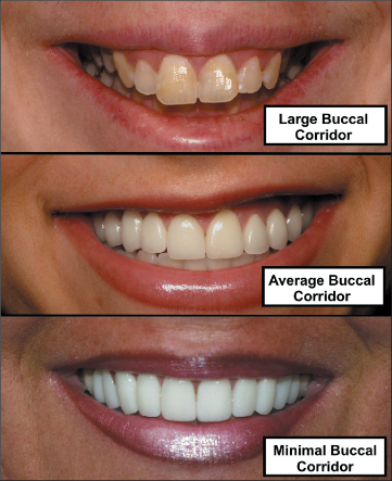
figure 15
Factors that Determine How Your Smile Looks

figure 16
figure 17
In others, the arch may be tapered or narrow creating huge triangular shadows. This creates a situation where little if any posterior teeth can be seen resulting in the illusion of hugh dark tunnels when this person smiles. The trian- gular shadows can be decreased by changing the arch form and width.
Dark Tunnels
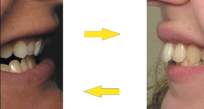
As we focus on the anterior teeth we see interesting tooth relationships between the maxil- lary and mandibular arches. When we examine the maxillary front teeth we notice that they can be in a normal relationship to the lower teeth, protrusive (too far forward) or intrusive (slanted in). (figure 17)
Protrusive
Intrusive
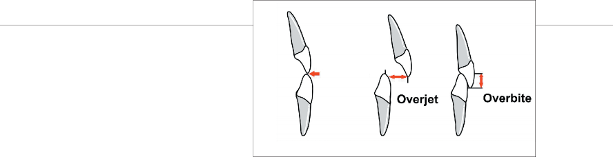
When anterior teeth overlap, this is referred to as an overbite. To be technically correct, spe- cific names are used when referring to overbite variations. When looking at a person’s smile from the side, the protrusion of the maxillary incisors beyond the edges of the mandibular incisors on horizontal plane is called the over jet. The amount that the maxillary incisors hang vertically down over the mandibular incisors is called the overbite. (figure 18)
These terms are often confusing. You can have an overjet with or without an overbite. Or you can have a deep overbite with great or little overjet.
CHAPTER TWO
Looking at the teeth from the frontal view, the six maxillary teeth have the greatest influence on the look of a smile. Therefore, special attention must be given to how the maxillary anterior teeth look and how they are arranged.
Anterior teeth come in many shapes and sizes.
When designing a new smile, each patient must first decide what shaped teeth are right for them. (figure 19)
Teeth can be square, tapered or ovoid. There can also be a combination of these shapes, such as square tapered or square ovoid. Several combinations of tooth shapes can be used with in an arch. A person can choose ovoid incisors and pointed canines. Or they could choose square and flat incisors and rounded canines. This is part of the artistic creative process that makes a particu- lar smile unique and attractive.
Edge to Edge
Overjet
Overbite
figure 18
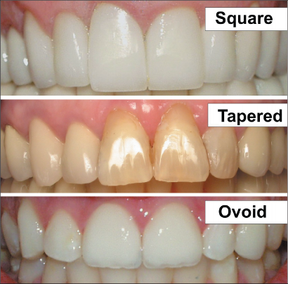
figure 19
Factors that Determine How Your Smile Looks
figure 20
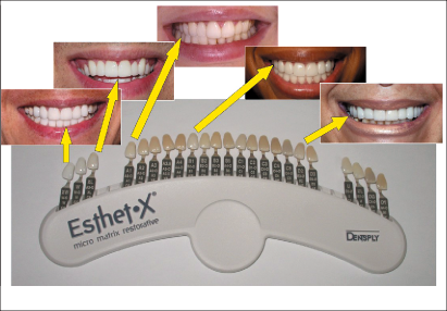
figure 21
Once the correct shape is selected, the focus should shift to how the teeth line up on a straight line (the horizontal plane), as viewed from the front of the patient. (figure 20). Remember, this horizontal plane should be parallel to the inter- pupilary line that connects the center of both eyes. There are many variations at the edges of the teeth, in relation to this line. The edges of the maxillary teeth (the incisal edges) may line up on a straight line, or they may be arranged so that the maxillary lateral incisors are shorter to varying degrees than the central incisors. Staggered incisal edges make a smile appear more natural and more youthful. Incisal edges on a flat plane make the edges look more worn. Worn edges are more commonly found in patients who grind their teeth or older patients as they wear down their teeth over time.
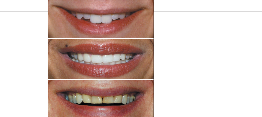
Color is another important consideration in the smile design process. There are dozens of tooth colors available. Generally speaking, it should be discussed with the cosmetic dentist whether a white smile look or a “natural” look is preferred. Teeth naturally are not very white, but have more cream-like or light yellow overtones. The specific colors of individual teeth can be cho- sen from shade ranges on dental shade guides. A cosmetic dentist can provide many shade guides from which to choose. (figure 21). For a more natural look, different color shades may be used within the same arch. For example canines are naturally darker than incisors or posterior teeth.
CHAPTER TWO

Choosing the correct color of teeth can be tricky. Given the same size teeth, the lighter the teeth are, the larger they will look. If teeth are more pronounced and forward in the arch form because of an individual’s specific anatomy, very light colored teeth should not be chosen. They will not look attractive. Instead, the color should be toned down. (figure21b). Similarly, when teeth are naturally very large or very long, very white colors should be avoided. On the contrary, if teeth are small or recessed in the mouth, lighter colored teeth will make the smile look better.
If any select teeth are being restored, it is important that the color chosen for these teeth blend in with the remaining teeth in the arch. This color does not necessarily have to be a perfect match if the other teeth don’t really show when smiling. However, they should be in the same range of color. If you are choosing to change the look of your front teeth, color will influence the complexity of your smile makeover. The lighter the color selected, the more teeth will have to be restored for the resulting smile to look natural and attractive. The average smile makeover requires a minimum of 8-10 maxillary teeth to be cosmeti- cally improved in order to attain a great looking smile. If the lower teeth show when you smile 6-
8 mandibular teeth may also have to be restored to compliment the look of the new maxillary teeth. If minimal mandibular teeth show when you smile, it may be enough to just bleach your lower teeth to make them brighter. (figure 21c)
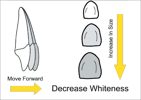
figure 21b
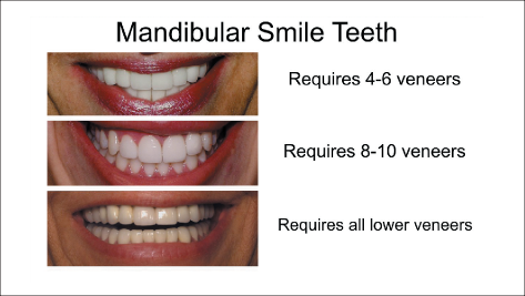
figure 21c
Factors that Determine How Your Smile Looks
Incisal Edge
MILD
MODERATE
HEAVY
figure 22
figure 23
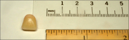
figure 24
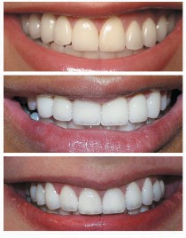
In addition to the colors, shapes, sizes and arrangements of teeth, the patient and the cos- metic dentist must also consider the biting edges of the anterior teeth. These biting edges, called the incisor edges, have a different property of enamel than the remaining tooth structure. (figure 22)

At the incisal edge, the enamel is usually more translucent than the thin enamel covering the the body of the tooth. This translucency allows light to easily pass through the incisal edge, giving the edge a blue-gray look.
This blue-gray look is caused by the back- drop and shadowing of the deepest part of the mouth showing through the translucent enamel. It should be decided by the cosmetic dentist and the patient if a mild, moderate or heavy amount of translucency is preferred.. (figure 23)
Another important fact to be aware of is how cosmetic dentists measure teeth. They measure tooth size in millimeters, not inches. Don’t refer to the customary inch scale when asking to have teeth lengthened, widened or changed. Millimeters may be very tiny, but adding just a millimeter can have a dramatic visual effect upon teeth and smiles. (figure 24)
CHAPTER TWO

Finally, no discussion about cosmetic dentistry would be complete without mentioning the “golden proportion”. The golden proportion was identified during the time of ancient Greece. When visualizing many objects in nature, it was discovered that there seemed to be a logical rela- tionship or constant proportion between those objects that could be measured. Interestingly, in mathematics and science, those same proportions could be found over and over again. (figure 25)
The golden proportion is a measurable and repititious pattern that is found throughout nature. When objects contain this golden propor- tion they are usually appealing. When two objects are in the golden proportion the larger object is 1.618 times the size of the smallest object. This relationship is measurable by a “golden propor- tion gauge”. (figure 25)
This formula can be seen in all of biology and throughout nature. Since it was discovered, it has been used by architects and designers throughout the ages. (figure 25b)
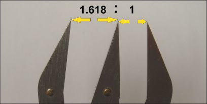
figure 25
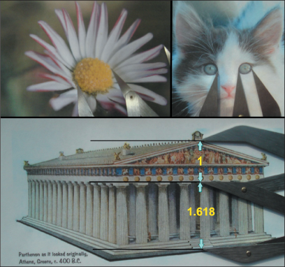
figure 25b
Factors that Determine How Your Smile Looks

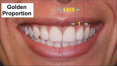
figure 26
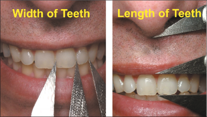
figure 26b
When teeth are arranged within the golden proportion, they appear visually attractive. Teeth are arranged in the golden proportion to each other as well as to other landmarks on the face. (figure 26) demonstrates how teeth are in a golden proportion to each other and now the length of teeth are in a golden proportion to the length of the upper lip. (figure 26b)
The golden proportion should be used as a guiding principle when creating esthetically beau- tiful teeth.
A basic understanding of dental anatomy will go a long way in effectively opening the channels of communication between patient and cosmetic dentist. A little knowledge of dental basics and guidelines allows patients to become a part of the creative process.
CHAPTER THREE

This chapter from Amazing Smiles Through Cosmetic Dentistry explains or demonstrates factors that determine how your smile looks as part of Dr. Albert J. Kurpis’s broader cosmetic dentistry approach to improving smile balance, appearance, and confidence.
The right treatment depends on the condition of the teeth, gums, bite, facial balance, smile goals, and a professional examination. Similar-looking smile problems may require different solutions.
Cosmetic dentistry is designed to improve the appearance of the teeth and smile. For many patients, a more balanced and attractive smile can also improve confidence in social, personal, and professional situations.
Use the related chapter links on this page to explore similar cosmetic dentistry cases, porcelain veneer examples, dental implant topics, bite correction cases, and full mouth reconstruction chapters.
Swipe left or right on mobile to move between book pages.